

A clever surgical technique can restore full function to the eyelid after growth removal, as Andrea Oakes discovers
SKIN masses, such as melanomas, sarcoids and squamous cell carcinomas, can be particularly troublesome when they appear in close proximity to the eye. Left untreated, a mass may grow and interfere with eyelid function or vision. Yet treatment can result in damage to the thin skin of the eyelid, inhibiting its ability to protect the eyeball and to keep it bathed in lubricating tears.
While nine-year-old Irish Draught/thoroughbred mare Rosie appeared unbothered by a cluster of ulcerated masses developing at the outer corner of her left eye, vets at Pool House Equine Clinic in Staffordshire felt that these growths were best removed.
“Treatments such as chemotherapy and radiotherapy are sometimes possible, depending on the nature of the mass, but in this case surgical excision was the favoured option,” says equine surgeon Marco Marcatili. “To preserve the eyelid anatomy and function, and to create a good cosmetic outcome, we planned a reconstructive technique called sliding ‘Z’ dermoplasty.”
DIGGING DEEP
ULTRASONOGRAPHY of Rosie’s eye revealed that the masses penetrated about a centimetre into the deeper tissue layers.
To minimise the likelihood of recurrence after excision, it would be necessary to remove
a significant amount of surrounding tissue during surgery. The aim of “sliding flap” reconstruction is to reconfigure what is left of the healthy tissue, to create less distortion of the skin and a neater scar.
The sliding “Z” flap is just one of a number of flap shape variations, explains Marco. Two triangular areas of skin adjacent to the surgical site are separated from the underlying tissue and removed. The remaining skin flaps are advanced (manoeuvred) into a zig-zag pattern, and the edges are then stitched together with absorbable suture material.
“The surgery is performed with the horse under general anaesthetic, as opposed to standing and sedated, because the surgical instruments are so close to the eye,” adds Marco, explaining that the slightest movement could result in injury.
“We always use ultrasound before surgery to evaluate the full extent of the mass and to plan the excision. Afterwards, the mass is sent to the lab for pathology.”
SEAMLESS REPAIR
FOLLOWING an uneventful recovery from the anaesthetic, Rosie was nursed at the hospital and given antibiotics and anti-inflammatories. Once it was clear that the site was healing without complication, she was allowed home.
“This technique can be very useful to close high-tension wounds following tissue removal,” says Marco, adding that lab tests identified the growths as sarcoids. “All surgery carries some risk, but we usually find that the eye area heals well and fast.”
Six months later, Rosie’s owner, Ellie-Mae Morson, reports that the surgical scar is barely visible.
“She had to wear a fly mask for a few weeks, to protect the area, but after a short time in the stable she could go back out to grass,” says Ellie, who regularly feels along the scar line for any signs of sarcoid reappearance. “Now the hair has grown back, you would hardly know it had happened.”
The sliding “z” flap: how it works
The periocular region (around the eye) is clipped and cleaned, and marked with incision lines.
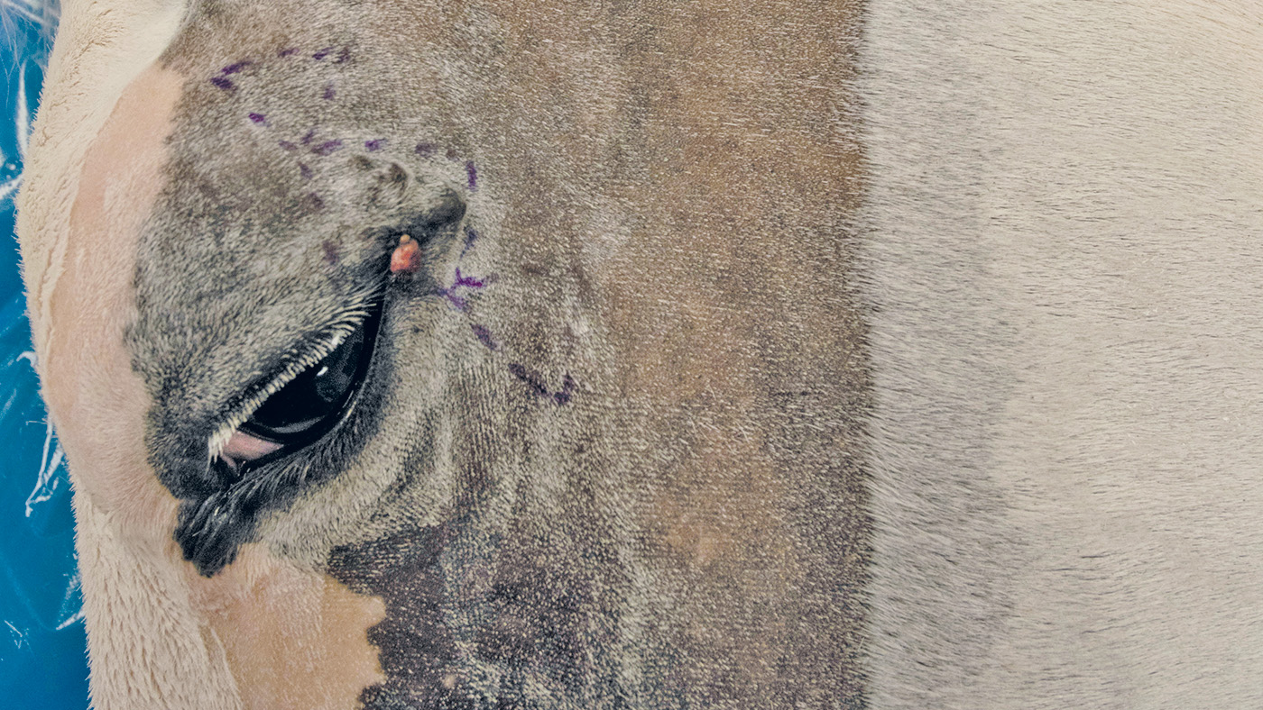
The masses are excised in their entirety, leaving an area devoid of skin.

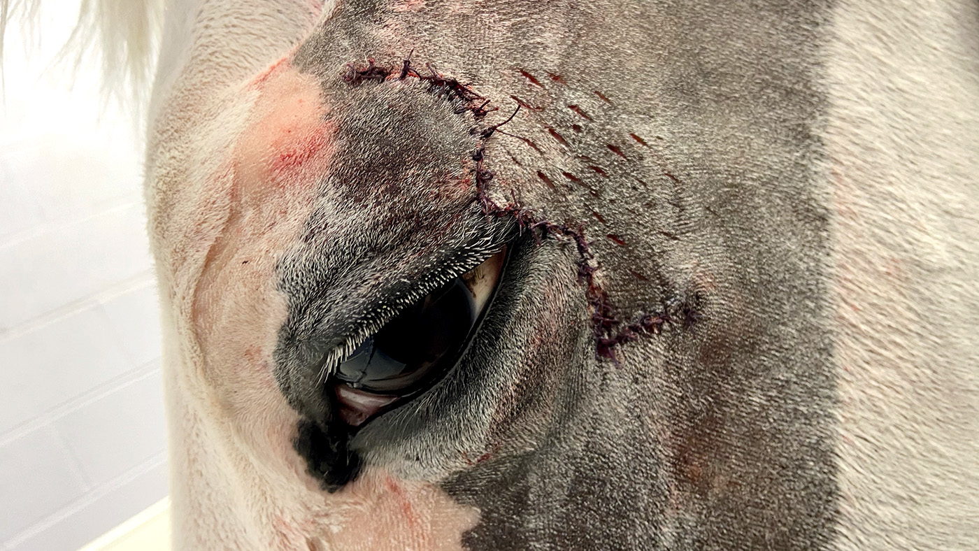
Adjacent triangles of skin are removed to let the remaining flaps be drawn across into place.

Suturing completes the reconstruction. Note the tension-relieving incisions behind the site.
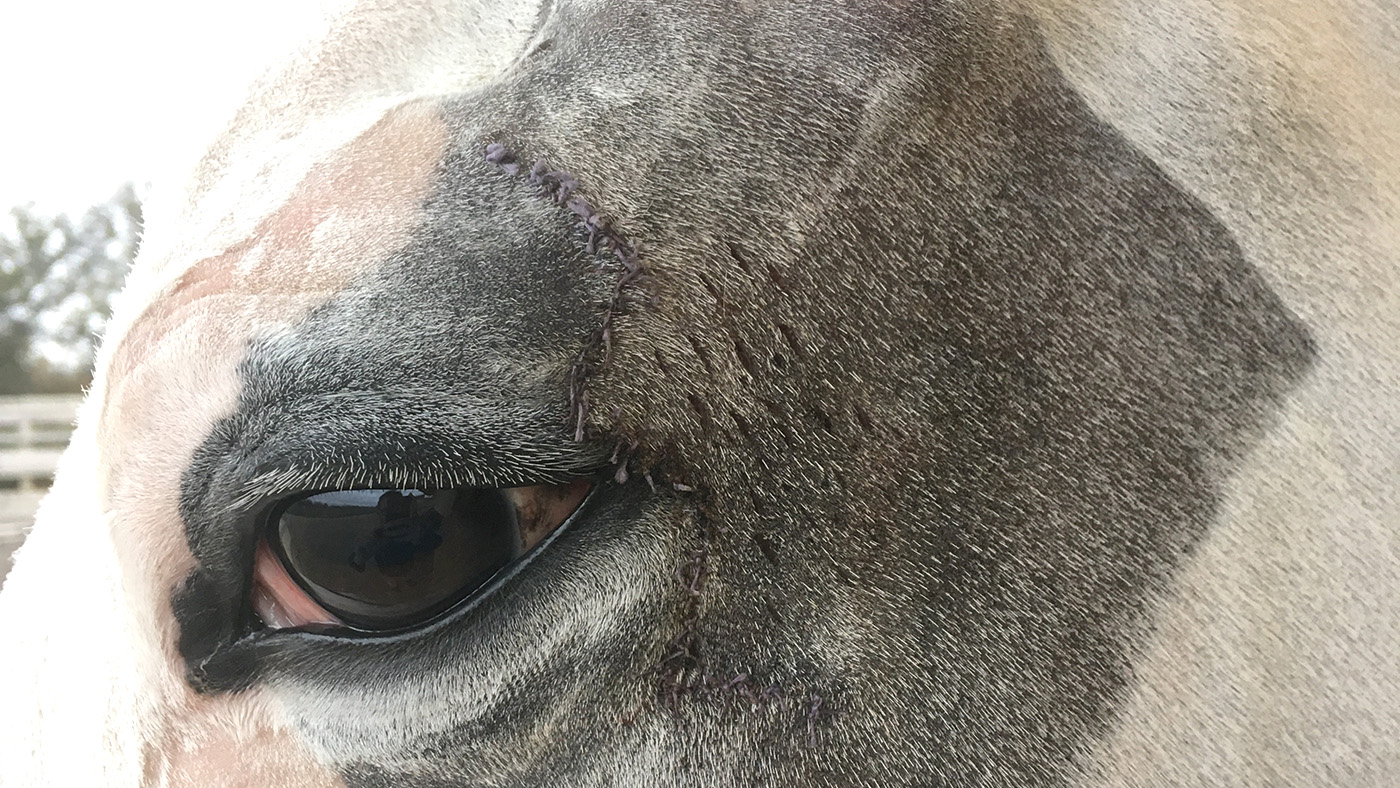
The eyelid is fully functioning and already beginning to heal, just 48 hours after surgery.
You can also read this feature in the 13 May issue of Horse & Hound magazine.
You might also be interested in…

Back conformation and saddle fit: what’s new in the veterinary world? *H&H Plus*
Play your part: how to help keep competition horses happy *H&H Plus*

How different grazing systems can help you control your horse’s weight *H&H Plus*

Keeping a veteran active for longer