
Poor performance or lameness with no obvious cause may call for an examination known as a work-up. Gil Riley MRCVS explains what’s involved
A lameness work-up is an investigation of the horse’s movement to diagnose injury or damage that is causing pain. The more accurate the diagnosis, the more finely tuned the resulting treatment plan can be. This, in turn, maximises the likelihood of a successful outcome and a return to athletic work in the shortest possible period of time.
A work-up may be performed because a reason for lameness is not apparent, or because performance is below par. There are many causes of loss of performance, including gastric ulcers, respiratory issues and temperament problems. By far the most common problem is orthopaedic pain, resulting from an abnormality somewhere in the musculoskeletal system.The aim of the work-up is to determine the root of this pain, using a sequential series of steps.
Where is the best place for a lameness work-up?
With so many unavoidable variables due to the individuality of every horse, it is important to standardise the procedure wherever possible.
A work-up is ideally performed at a veterinary clinic, where the vet will be familiar with how horses respond to the environment, the handlers and the facilities. These facilities will have been designed to allow the most obvious demonstration of lameness in the horse in the safest way — a small gravel lungeing ring, for example, that enables the horse to be seen moving on a hard surface with minimal risk of slipping.
Typically, one vet will use their experience and the examination results to lead them to the source of pain. A good lameness clinician needs to ensure they remain sufficiently objective to pinpoint the diagnosis. An advantage of bringing a horse to an equine hospital is that there may be more than one specialist, along with gait analysis systems and different imaging techniques that may be unavailable at home.
In certain cases, variations to the procedure may be necessary. A horse that has recently had its shoes removed may not be lunged on a hard surface, to avoid making him footsore, while the riding part would be omitted, of course, for an unbroken or miniature horse.
If the answer is found with the initial, static examination, or on the straight-line trot, the process ends there. It would be inappropriate to put the horse through unnecessary discomfort that would provide no further information.
If a horse will not tolerate nerve-blocking injections, we may opt to introduce imaging earlier than usual to minimise distress caused. This is not ideal, as it will take more time to reach the correct diagnosis, but it is a better alternative to risking injury to the horse, vet or handler.
Unravelling issues
Lameness in the limbs is most common, but pain in other areas can result in poor performance — especially back issues, such as impingement of the dorsal spinous processes (kissing spines) or spondylosis of the vertebrae. Such horses often exhibit no lameness on the straight-line trot or on the lunge, but show profound discomfort during ridden exercise under the weight of tack and rider. They may also resent palpation of the back, alerting the vet to the possibility of a problem in this area.
A work-up can also help to unravel a primary source of pain from secondary complications or compensatory lameness. An example would be hind limb suspensory pain — a common cause of lameness in dressage and event horses. Because it can develop insidiously, and in both limbs simultaneously, the rider may not realise anything is wrong in the early stages. The horse may adapt his movement to minimise pressure on the painful suspensories by moving his pelvis more, causing sacroiliac pain. When his suspensory ligaments are blocked out (numbed), the clinic rider is likely to report that he now moves more freely — but with a stiffness “higher up”. A subsequent nerve block of the sacroiliac area will then lead to further improvement.
Pain relief, such as phenylbutazone, should not be given for at least two days before the exam. Anything that makes lameness less obvious will make obtaining an accurate diagnosis more difficult.
What’s the verdict?
Eliminating possibilities to reach the correct diagnosis can take money and time.
While a definitive diagnosis is obtained in most cases, some may still evade an absolute answer — although a logical and well-supported suggestion can usually be made. Lamenesses that remain undiagnosed are usually suspected as being neurological in origin, emanating from the spinal cord or the associated spinal nerves and plexuses.
The emergence of gait analysis systems has been an exciting development in lameness investigation, particularly for horses that are unbroken or have become unrideable, or where lameness is so subtle that it is difficult for the vet to identify. These systems, however, should be regarded as another tool available to the vet, and just part of the process in uncovering the source of some lamenesses.
A lameness work-up: the process in detail
Part 1: history
The horse’s background is explored thoroughly, from specifics of ownership and use to any previous lameness issues or suspicions. Other health conditions are also noted, along with the horse’s shoeing regime, the nature and onset date of the current lameness, details of any treatment and subsequent response.
Part 2: static exam
Each limb is palpated for swellings, heat, bony lumps or signs of traumatic injury, along with response to pressure. The feet are inspected, with attention paid to shoe fitting, the presence of a pulse and any pain upon use of the hoof testers.
The back and neck are palpated for areas of sensitivity or exaggerated pain reaction, and baited (carrot) stretch tests performed to assess neck manoeuvrability.
Part 3: dynamic exam
A. Straight-line trot: Unless severe lameness is already evident at walk, the horse is trotted away and back on a hard, level surface. As a two-time gait, trot allows direct comparison between sides. All but the mildest cases of lameness are usually apparent. Flexion tests may be performed at this stage. Any change in gait in the trot-away may point to a problem in a joint or an associated structure.
b. Hard-surface lunge: lungeing on a tight circle can provide vital information. Lameness that is particularly obvious on a hard surface may stem from a foot. Noting whether it worsens when the affected limb is on the inside or outside can help to pinpoint the pain source to a certain area of the foot.
c. Soft-surface lunge: lameness that is worse on the soft indicates that the problem is unlikely to be in the foot and may instead be in a soft tissue structure, such as a ligament or tendon. Further clues can be gained if it worsens in one direction.
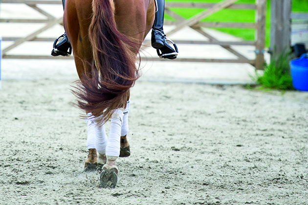
d. Ridden exercise: the majority of lameness work-ups are brought to us by owners not because a horse looks wrong, but because he feels wrong.
An experienced rider can be invaluable in helping the vet determine the source of this abnormal movement, or lameness, by reporting, for instance, that the horse feels choppy on both front limbs, is not tracking up with a hindleg or resents pressure on his back.
Part 4: nerve blocks
Possibly the most important tool in lameness diagnosis and often the cornerstone of the investigation. While “pain” cannot be imaged, the vet can narrow down the problem by progressively blocking out regions of the affected limb, or spine, before re-assessing movement. If an area is no longer painful, a horse’s gait will improve significantly — indeed, he may become sound. Such a response will categorically regionalise the source of pain.
Part 5: imaging
With the source of pain localised, imaging can be employed to assess the specific area — commonly radiography, ultrasonography, MRI (magnetic resonance imaging) and nuclear scintigraphy. Using information about the damage found, along with knowledge and experience of dealing with lameness, the vet may reach a diagnosis and formulate a treatment plan.
This feature was first published in Horse & Hound magazine; 9 January 2020
You might also be interested in:

Lameness in horses: what every owner needs to know

How blocking a horse’s nerve or joint can help locate a source of pain
Suspensory ligament injuries: what owners need to know

Kissing spines in horses: what all owners need to know

Subscribe to Horse & Hound this spring for great savings
Horse & Hound magazine, out every Thursday, is packed with all the latest news and reports, as well as interviews, specials, nostalgia, vet and training advice. Find how you can enjoy the magazine delivered to your door every week, plus options to upgrade your subscription to access our online service that brings you breaking news and reports as well as other benefits.