

While a lump or bump is never welcome in the showing world, a prominent growth on the shoulder of Our Cashel Blue took a particularly sinister turn last September. Tests on the lump, which was becoming larger and more ulcerated, revealed a rare form of cancer — devastating news for connections of the usually hale and hearty champion cob.
When his owner Caroline Tyrrell and the Hood Show Team announced his impending operation on Facebook, Blue’s fans responded with a collective “fingers crossed” for successful surgery. He is now home and recuperating at the Hood’s yard after removal of the tumour, but is he completely in the clear?
Suspicious signs
Blue’s lump was situated on his right side, in the large infraspinatus muscle that lies on top of the scapula (shoulder blade).
“As a horse vet, I see a lot of lumps,” says James Tilly MRCVS, of the Hoods’ veterinary practice Knotts Yard. “Most are sarcoids, but Blue’s didn’t have that look. It was ulcerated on the surface and quite sore.
“Sarcoids in horses usually form in areas such as the sheath, inner thigh or eyelid,” adds James. “This lump didn’t fall into any of the typical sarcoid categories — it seemed softer and more embedded in the underlying muscle.”
James performed a biopsy called a fine needle aspirate to extract tissue for analysis.
“Taking a sample can make the growth angry and aggressive, but we needed to find out more,” he explains.
The samples were sent to the Royal Veterinary College (RVC), where cytology (microscope analysis) identified the presence of mast cells. These cells, produced in the spongy bone marrow found in the centre of larger bones, help fight infection as part of the immune system. In this case, the cells had undergone a transformation to create a cancerous tumour.
A mast cell tumour forms within the skin (cutaneous) or just beneath it (subcutaneous). Andy Fiske-Jackson MRCVS, a surgeon at the RVC, says: “Mast cell tumours are uncommon in horses, representing around 7% of all skin tumours. The cause is unknown and there is no strong breed or gender predilection. They’re not usually malignant and rarely spread between organs, so we could be a bit more confident that complete removal of the tumour would be curative.
“The lump was too large to remove at the yard, so we decided on surgical excision at the RVC Equine Referral Hospital.”
The aim was to remove the 7cm tumour in its entirety, along with sufficient surrounding tissue, to minimise chances that any of the cancer cells would remain and regrow or spread.
“The infraspinatus muscle is important for shoulder stability,” says Andy. “The distinction between a tumour and the thickened skin around it is not always clear. While we didn’t want to leave a hole so big that we couldn’t close it, we had to take enough of a margin either side of the tumour as well as beneath it.
“The shoulder is a high-motion area, so we were concerned that the wound might break down,” he adds “As Blue is a show horse, cosmetic appearance was also high on the agenda. Matching up his coat markings during skin closure was quite tricky.”
Andy employed a variety of stitching techniques to bring the edges of the 10cm hole together, using intradermal (embedded) and absorbable sutures for minimal scarring.
“Surgery went well,” says Andy. “Mast cells can release histamine when surgically manipulated, causing localised itching and suture rubbing, so we administered steroids pre-operatively. Blue was also given antibiotics and painkillers.”
Further tests
The concern with a cancerous tumour is metastasis — the development of secondary malignant growths at a distance from the primary cancer site.
“Blue is a big horse, so searching for metastases to a lump would be challenging,” explains Andy. “The lungs would be the first place to look: cells released by the tumour enter the bloodstream and travel to the heart; the heart then sends blood to the lungs, where the cancer cells become lodged in the small vessels. Rarely, cancer can also spread to the local lymph nodes and the abdominal cavity.
“While you can X-ray the lungs of a foal or a thoroughbred to a point, it’s impossible with a larger horse — especially with sufficient sensitivity to see small cancerous masses. Nor can you see into the lungs with an ultrasound scan, because ultrasound can’t travel through air.”
Laboratory tests suggested that the tumour was non-aggressive and unlikely to spread.
“We knew from the excision that we had good margins,” says Andy. “Lab results revealed a low mitotic rate, which meant cells were not dividing rapidly as in the case of an aggressive tumour. Had it been a malignant type we would have been in a difficult place — we would have expected cancer to occur elsewhere.”
Is chemotherapy an option for equine cancer?
“We don’t tend to use systemic chemotherapy with intravenous drugs,” explains Andy. “The cost is prohibitive, due to the sheer quantity of drugs required, and there are ethical questions about side effects — the idea of making a horse very sick with a treatment with no known outcome.
“We use local chemotherapy, however, for conditions such as sarcoids and melanomas,” he adds. “Injecting drugs into these skin-based or subcutaneous tumours is feasible and it’s something we’re really moving forwards with.”
Thankfully, Blue’s rare tumour was treatable. Complacency with any lump, bump or lesion, however, is ill advised.
“It could well be a harmless fibrous lump or an insect bite, but we see a lot here that have become difficult,” he says. “While there has typically been a ‘wait and see’ policy, it is always worth getting a diagnosis earlier rather than later. A smaller lump is easier and, usually, cheaper to deal with.
“If a wound or a sore is not healing well, ask yourself why,” adds Andy. “Often, there’s an underlying tumour or sarcoid.
“By knowing what the problem is, you and your vet can make an informed decision about treatment. If you leave it too late, the decision may already be made for you.”
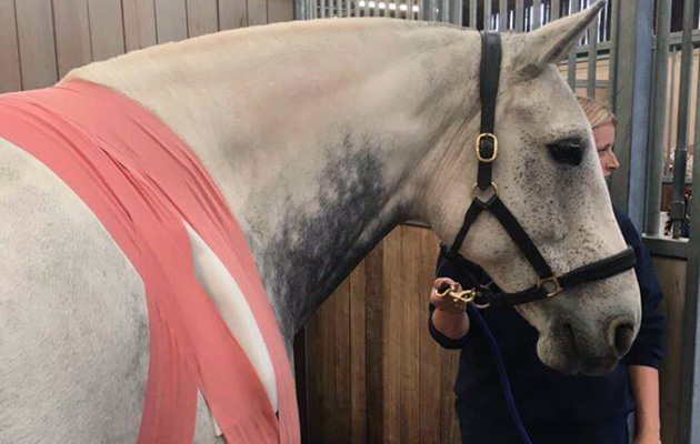
Progress report
Following surgery, the biggest challenge was keeping the dressing in place — hence the convoluted bandage arrangement over Blue’s chest and withers. It was then a case of monitoring the nine-year-old gelding for problems with healing and signs of recurrence.
Allister Hood, who has ridden Blue to major victories at the Horse of the Year Show and Royal International, says: “We’re taking good care of him and giving the shoulder time to heal. He was confined to his box at first, as we didn’t want to turn him out and damage the wound or set back the healing process. We then led him out for a few minutes for a pick of grass, before gradually building up the walking.
“It will be a long winter for Blue but he has been incredible, bless him,” adds Allister. “At the moment, signs are positive.”
Ref Horse & Hound; 3 January 2019